Research Article | | Peer-Reviewed

Identification of Yak Hair Scanning Electron Microscopy in Northwest China

Received: 2 April 2025     Accepted: 15 April 2025     Published: 9 May 2025
Views:       Downloads:
Abstract

Yak (Bos grunniens) was a unique species living in the Qinghai-Tibet Plateau and grassland which has plateau cold climate. With excellent warm, soft, breathable features, yak hair has always been woven as raw materials for the tent by local herders. China's yak hair resources were very rich, with an annual output of yak hair up to 10,000 tons or more. Yak felt products were expensive. Criminals used the hair of other animals to impersonate yaks and made felt for sale. This behavior disturbed the order of the market and damaged the legitimate rights and interests of consumers. In order to understand the characteristics of yak hair, this paper observed and analyzed the different of yak wool fibers between Tibet and Qinghai. The fresh yak hairs of Tibet were used as the experimental group and the yak specimens of Qinghai were used as the control group. The crude fibers of Yak leg were a myelinated hair and consisted of the scale layer, the cortex and the medulla layer by SEM (scanning electron microscopy). The wear degree of coarse wool fiber at the same parts was lower than the two-type hair fiber. There were differences in the structure of scales between black and white hairs. The characteristics of yak hair were very obvious, and the microstructure differences of yak hair between different production areas were also significant. The electron microscope detection operation was simple and the results were clear. This method can be used as a rapid identification technique for identifying yak felt products.

Published in Animal and Veterinary Sciences (Volume 13, Issue 2)
DOI 10.11648/j.avs.20251302.12
Page(s) 45-51
Creative Commons

This is an Open Access article, distributed under the terms of the Creative Commons Attribution 4.0 International License (http://creativecommons.org/licenses/by/4.0/), which permits unrestricted use, distribution and reproduction in any medium or format, provided the original work is properly cited.

Copyright

Copyright © The Author(s), 2025. Published by Science Publishing Group

Keywords

Yak, Scanning Electron Microscope, Hair Characteristics, Tibet, Qinghai

1. Introduction
Yaks are distributed in the alpine meadow vegetation belt of Xizang, Qinghai, Sichuan, Xinjiang and Gansu provinces in China, and are concentrated in the high altitude area of 3000~6000 meters above sea level . Yaks are a typical high-altitude animal that is extremely cold resistant. It can survive under special conditions of cold and humidity, which is related to the unique structure of its hair . Through SEM (SEM is an abbreviations of the scanning electron microscopy), it was found that the scale layer on the surface of yak hair is tightly arranged and irregularly wavy, with a well-developed medulla layer . These structures help enhance the insulation, waterproofing, and UV resistance of the hair, adapting to high-altitude and hypoxic environments.
The microstructure of yak hair fiber mainly depends on the innate genetics of the animal and has species specificity. The formation of yak hair color is related to gene expression . There are three main structures of animal hair fibers, from outer to inner, which are the scale layer, cortex layer, and medulla layer. The scale pattern of the scale layer and the medulla pattern of the medulla layer have classification functions and can be used to distinguish different types of animal hair. At present, animal classification is mainly based on scale morphology, medullary pattern, pigment granule morphology, and their distribution . There are studies that use the differences in microstructure of yak hair to distinguish between wild yaks and domestic Yaks . But currently, there is no use of yak hair to distinguish yaks based on gender differences, hair color changes, or different geographical regions.
Yak hair can be divided into three types: long soft hair, Tow-type hair, and coarse hair. Liu found through micro mechanical testing that the tensile strength of yak wool fibers is better than that of ordinary wool, but the scale structure makes its products prone to shrinkage and requires chemical modification treatment . Yak felt products are products made by modifying hair. But because of the poor spin ability, large static electricity and complex production process of yak wool fiber , yak felt products were expensive. Due to its high profits, there have been numerous cases of counterfeit yak felt products, seriously disrupting the order of commerce and trade. Although DNA technology can identify species, it is difficult to extract genetic material from chemically treated yak hair using DNA technology. Moreover, microscopic examination is more cost-effective than DNA identification and is more easily accepted by businesses. Therefore, SEM has become the most cost-effective method for species identification. In this study, SEM was used to analyze the differences between the characteristics of yak coarse hair fiber and Tow-type hair fiber, and the microstructure characteristics of Tibet yak and Qinghai yak hair fiber were compared, in order to provide technical support for the classification and forensic identification of yak hair fiber. .
2. Materials and Methods
2.1. The Experimental Animal Hair
The yak hairs were included coarse hair fiber and type II hair. These coarse hairs and two-type hairs were divided into black and white types by hair color. This research took no account of the age and sex-related difference. The comparative experiment between one control group (a yak specimen) and one experimental group (a yak) was conducted.
Three body parts of yak, head, trunk and leg were chose, and random designed the quadratsin every part.10 coarse fibers were selected in each quadrat, in total 30 coarse fibers were extracted. Then 10 two-type hairs were extracted from trunk. The two-type hairs just were compared with the coarse fibers.
The control group hairs were collected from Nanjing Police University specimen room. The yak specimen came from Qinghai Province. The control group hairs included the black coarse fibers, derived from the yak head, trunk and leg respectively; and the black two-type hairs derived from the trunk.
The experimental group yak hairs were collected from the pasturing area in Tibet. The experimental samples were difficult to obtain, and the collection amount and location were limited, because of the yaks were aggressive and difficult to approach. The black and white coarse fibers just derived from the yak leg. Only 10 black coarse fibers of yak leg were compared with the control group. Then 10 white coarse fibers just were compared with the black coarse fibers.
2.2. Methods
After stoving the samples, put in 95% ethanol: ethyl ether (1:1) and degreased for 20min. The defatting samples were washed in anhydrous ethanol for 2min and then dried with filter paper . The samples were fixed on the sample table with double-sided tape, and under the s-4000 type scanning electron microscope (SEM), the morphology of the hair was observed and photographed. The acceleration voltage was 10KV.
3. Results
All hairs had been examined by SEM, showed that the ultrastructure of hairs was made up of scale layer, cortical layer and medulla layer. The scale layers located in the outermost layer were very thin, the main protection. The cortical layers by degradation of cells arranged in a disorderly density of homogeneous.
3.1. The Experimental Group (Tibet)
3.1.1. SEM of Dark-haired Fibers in the Leg
The SEM photographs of yak legs black coarse fibers showed that the dark coarse fibers had narrow apexes, the width was half of the middle, larger scales and loosely arranged (Figure 1). Central hair fiber diameter larger, scales closely packed, the pattern were messy imbricate. The scales widths were less more than 11 microns. The root hair follicles were filamentous and wrap around the scales. Peel the root hair follicle to see the medullary layer. The medulla of the medullary layer was dense, well-arranged and had smooth edges. The medullary layer of the hair was composed of a gas chamber and a chamber wall.
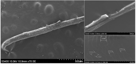
Figure 1. Hair follicle, the medulla layer (Hair follicle longitudinal section) and the scale width of scale layer.
3.1.2. SEM of White-haired Fibers of the Leg
The SEM photographs of white coarse fibers showed that the white hair cortical layer was near the hair side and had a white matter, which was assumed to be a white area that was absent from melanin (Figure 2). From the tip to the root of scale layer, scaling change from loose to close, scales angle became smaller, scales width also gradually decreases, and more than 10 microns, like a fish scales closely pasted on the hair shaft.
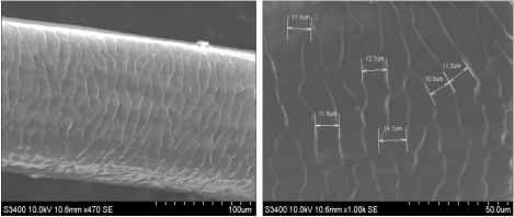
Figure 2. The pattern and width of the scale layer of hair shaft.
Table 1. Scale structure of dark and white coarse fibers of yak legs in Tibet.

Sample

Scale type

Sorting order (from hair tip to root)

dark coarse fibers

Coronal, Clutter type, Petal type, Flat type

Clutter type→Flat type

white coarse fibers

Clutter type, Petal type, Flat type

Petal type→Flat type

Note: Classification reference for scale patterns .
Black and white coarse fiber scales of yak all had flat type. Among them, dark-colored coarse fibers were mainly clutter type, and white coarse fibers were mainly composed of hybrid type flaps (Table 1). There were obvious coronal scales on the tip of the black coarse hair fibers, and the middle part of the hair was mainly clutter type, and gradually was flat type near the hair root. No coarse coronal scales were found in the white coarse fibers. The scales were petal type, and the others were similar to the black coarse fibers. The results showed that the scale types of the coarse hair fibers of the two color types were basically similar and the differences were not significant.
3.2. The Control Group (Yak Specimen of Qinghai)
3.2.1. SEM of Dark-haired Fibers of the Head
The scale layers of the hair of yak samples were more severe crack (Figure 3). The root and central severely damaged, the tip of hair little damaged. The scales on both sides of the fiber were superior to those of the middle of the fiber, and the middle scales were blurred. Compared with the coarse hair fibers of the experimental group yaks, the scales of yak specimens were damaged, and were scraped away. The posterior cortical layer was similar, without obvious damage.
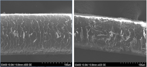
Figure 3. Damaged scale and Cracked scales.
Compared with the control group and the experimental group, the dark coarse fibers of the legs, although the scale layer of the yak specimens were severely damaged, the cortical layer structure was similar and was not significantly damaged. It showed that the damaged scales of yak legs hair could still played a protective role. The lamellae were composed of keratin proteins that act to protect cortical cells.
Yak's long hairs were mainly dominated by coarse fibers. The head fibers scale damage was obvious, but the scales were still clearly visible, and the scale pattern was dominated by the clutter type. The scales were large and closely aligned, and the area of the lesion was warped. Some of the scales were badly damaged and cannot be identified. The rift was severely split.
3.2.2. SEM of Dark-haired Fibers in the Leg and Trunk
The leg fibers scales layers were in a form of irregular, and severely damaged. The leg fibers scales layers showed that the coarse fibers of the torso were more damaged than the head, and the scales were almost invisible (Figure 4). The thickness of the fibers decreases after the scale layer was damaged, and the cortical and medullary layers were faintly visible, and the scales were more or less clear.
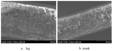
Figure 4. The scale layer of the hair shaft.
Table 2. Comparison of scale patterns of dark coarse fibers in yak specimens.

Body part

Scale type

Scale damage

head

Clutter type

Flake cracks more, showing cleavage.

leg

Flat type

Hairy central scales had a greater degree of wear and less scale on both sides.

trunk

Flat type

Scales had more warping angles, and the degree of wear was larger, and most of the scales were blurred.

The wear degree of the scale layers of different body parts under same storage conditions was different (Table 2). Severe abrasion happened in the scale layer of head coarse fibers. The scales on the tip of the hair were worn less, the middle part and the root appear split, and even the cortical and medullary layers were visible. The scale layer hardness of the fibers tip was greater than the middle and root of fibers. The trunk and leg hairs were well preserved and no severe scale cracking occurs. The scale layer of the coarse fibers tip of the leg was less damage, and the integrity of the scale layer on both sides of the fiber were better than that of the middle portion.
3.2.3. The SEM of the Two-type Hair Fibers
The damage of the two-type hair fibers from the tip to the root gradually increases. The scales of the hair tips were clearly visible, and the scales were large and closely aligned, with a coronal or hetero-wave pattern, and the wear scales had a warping phenomenon, and the scales were about 3~6μm. The two-type hair was dominated by clutter, and there was no medullary layer (Figure 5).
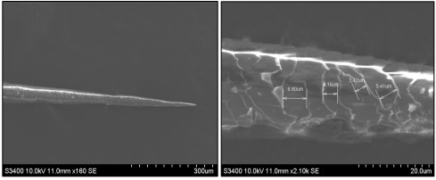
Figure 5. The general and scale of the two-type hair.
White material appeared in the middle of the hair, near the cortical layer, and the white material in the hair root spreads to the entire cortical layer. There was a fracture in the two damaged fibers, and the white material was broken and the scales were hidden (Figures 6~7). Tow-type fibers were more vulnerable to damage than coarse wool fibers (Table 3).
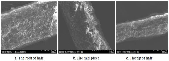
Figure 6. The detail pictures of the type II hair (a, b, c).
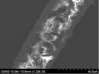
Figure 7. Scales of cracked phenomenon.
Table 3. Comparison between coarse hair and two-type hair scales of yak specimens.

Sample

Scale type

Scale damage

coarse hair

Flat type

Scales had more warping angles, and the degree of wear was larger, and most of the scales were blurred

two-type hair

Clutter type

The scales on the tip are lightly damaged, and the scale layers in the middle and roots are heavily worn, with white substances.

4. Discussion
Through SEM observation, the crude fibers of yaks are composed of scale layer, cortex layer, and medulla layer. Natural pigments were stored in the cortex. The formation of yak hair color is related to gene expression . The uniform arrangement of pigment particles along the hair shaft axis is the main factor affecting hair color. The scale structure does not affect the distribution of pigment particles . The whitening status of yak hair is mainly related to the distribution of pigments in the cortical layer. The structure of the medullary layer is related to the type of hair and does not affect pigment precipitation.
According to Table 1 and Table 2, there are differences in the microstructure of the scale layer of yak hair between Xizang and Qinghai. Therefore, the scale layer structure of yak hair can serve as a classification basis for distinguishing different distribution areas. In addition, the degree of hair damage in different parts of the body is also a significant interfering factor that cannot be ignored. The measurement elements such as scale arrangement order, scale density, and width can be important indicators for yak identification .
The special structure of yak hair allows them to survive in cold and humid environments. The two types of hair of yaks lack a medullary layer, and the scale layer has strong anti-corrosion properties. The reasonable arrangement and density of scales are the result of yaks' long-term adaptation to high-altitude living conditions.
5. Conclusion
As an independent species, yak hair has unique recognition characteristics, which is different from other Bovidae . The difference of microstructure of yak hair has certain taxonomic significance. According to the results of this study, there are significant differences in the microstructure of yak hair in different distribution areas of the same species, Tibet and Qinghai. Therefore, the microstructure of yak hair can be used as the identification basis between different species and different distribution areas of the same species.
Abbreviations

SEM

Scanning Electron Microscopy

Acknowledgments
This work was supported by the Special Funds for Basic Scientific Research Services of Central Universities (No. LGYB202505), and National key R&D Projects (No.2023YFC3304000), and Research Project of Higher Education Reform in Jiangsu Province (No.2021JSJG292).
Author Contributions
Yilin Li is the sole author. The author read and approved the final manuscript.
Conflicts of Interest
The authors declare no conflicts of interest.
References
[1] Zhong Jincheng, 2000. Chen Zhihua. Research on genetics and breeding of Yak [J]. Yunnan Journal of Animal Science and Veterinary Medicine, 2: 15-17.
[2] Li Hongmei, Yang Lingping, Bao Yongqing, et al.. 2024. Analysis of Wool Quality of Meiren Yak [J]. Journal of Animal Science and Veterinary Medicine, 43(6): 25-28.
[3] Shen Yushuang, Li Jibiao, Shi Yonghong. 2003. Scanning electron microscope observation of yak hair [J]. Journal of electron microscopy, 6: 490-491.
[4] Shi Lin, GUO Zi-Xuan, LI Xiao-Wei, et al. 2025. Cloning and Tissue Expression Analysis of Coat Color Candidate GeneSTX17 in Maiwa Yak (Bos grunniens) [J]. Journal of Agricultural Biotechnology, 33(4): 802-813.
[5] Hou Senlin, Xue Xiaoming, Song Qingshuang. 2012. Scanning electron microscopy analysis of the straight hairs of Mustela sibirica and Mustela erminea [J]. Nanjing Forestry University Journal (Natural Science Edition), 4: 149-152.
[6] Wang Liangjing, Hou Senlin. 2020. An observation on the hair of wild Yak and domestic Yak by SEM [J]. Journal of Jiangsu Forestry Science & Technology, 47(01): 49-52.
[7] Liu Jian. Research on the Morphological Structure and Stretching Refinement Process and Mechanism of Yak Coarse Hair [D]. Donghua University, 2010.
[8] Ren Yu, Chen Lian, Guo Xiaodong, et al.. 2025. Development of Cotton Spinning Equipment Spinning Coarse Count Pure Yak Wool Products [J]. Textile science and technology progress, 47(3): 39-42.
[9] Li Ming, Liu Jianwei, Hu Lichao, et al.. 2025. Discussion on dyeing and finishing process of hemp/organic cotton/yak cashmere corduroy [J]. Textile Dyeing and Finishing Journal, 47(01): 13-16.
[10] An Guoce. 2024. Identification of animal fiber by scanning electron microscope and infrared spectroscopy [J]. Wool Tetile Journal, 52(09): 111-116.
[11] Hou Senlin. 2010. Scanning Electron Microscope Analysis of 6 Kinds of Canine Straight Hairs in China [J]. Journal of Anhui Agricultural University, 4: 627-630.
[12] Sun Zhongwu, Gao Haiyu, Bi Bing, Wang Hongwei. 2003. Scanning electron microscopy analysis of deer animal hairs[J]. Journal of Northeast Forestry University. 4: 29-32.
[13] Wang Caiyun, Tian Xiaorui. 2009. Morphological structure and identification of several animal fibers [J]. Quality and Technical Supervision Research, 1: 26-29.
[14] Ma Yuhong, Ma Shike, Sun Wu, et al.. 2024. Analysis of Wool Fiber Quality of Plateau Type Tibetan Sheep Carpetin Different Areas of Qinghai Province [J]. Chinese Qinghai Journal of Animal and Veterinary Sciences, 54(04): 48-49+64.
[15] Wang Yuzhu, Zhu Hanqi, Luo Hao, et al.. 2022. Morphological Structure and Tensile Test Evaluation of Yak Cashmere Fiber [J]. Journal of Textile Science and Engineering, 39(02): 21-24+33.
Cite This Article
  • APA Style

    Li, Y., Lin, P., Xue, X., Zhou, S. (2025). Identification of Yak Hair Scanning Electron Microscopy in Northwest China. Animal and Veterinary Sciences, 13(2), 45-51. https://doi.org/10.11648/j.avs.20251302.12

    Copy | Download

    ACS Style

    Li, Y.; Lin, P.; Xue, X.; Zhou, S. Identification of Yak Hair Scanning Electron Microscopy in Northwest China. Anim. Vet. Sci. 2025, 13(2), 45-51. doi: 10.11648/j.avs.20251302.12

    Copy | Download

    AMA Style

    Li Y, Lin P, Xue X, Zhou S. Identification of Yak Hair Scanning Electron Microscopy in Northwest China. Anim Vet Sci. 2025;13(2):45-51. doi: 10.11648/j.avs.20251302.12

    Copy | Download

  • @article{10.11648/j.avs.20251302.12,
      author = {Yilin Li and Ping Lin and Xiaoming Xue and Shu Zhou},
      title = {Identification of Yak Hair Scanning Electron Microscopy in Northwest China
    },
      journal = {Animal and Veterinary Sciences},
      volume = {13},
      number = {2},
      pages = {45-51},
      doi = {10.11648/j.avs.20251302.12},
      url = {https://doi.org/10.11648/j.avs.20251302.12},
      eprint = {https://article.sciencepublishinggroup.com/pdf/10.11648.j.avs.20251302.12},
      abstract = {Yak (Bos grunniens) was a unique species living in the Qinghai-Tibet Plateau and grassland which has plateau cold climate. With excellent warm, soft, breathable features, yak hair has always been woven as raw materials for the tent by local herders. China's yak hair resources were very rich, with an annual output of yak hair up to 10,000 tons or more. Yak felt products were expensive. Criminals used the hair of other animals to impersonate yaks and made felt for sale. This behavior disturbed the order of the market and damaged the legitimate rights and interests of consumers. In order to understand the characteristics of yak hair, this paper observed and analyzed the different of yak wool fibers between Tibet and Qinghai. The fresh yak hairs of Tibet were used as the experimental group and the yak specimens of Qinghai were used as the control group. The crude fibers of Yak leg were a myelinated hair and consisted of the scale layer, the cortex and the medulla layer by SEM (scanning electron microscopy). The wear degree of coarse wool fiber at the same parts was lower than the two-type hair fiber. There were differences in the structure of scales between black and white hairs. The characteristics of yak hair were very obvious, and the microstructure differences of yak hair between different production areas were also significant. The electron microscope detection operation was simple and the results were clear. This method can be used as a rapid identification technique for identifying yak felt products.
    },
     year = {2025}
    }
    

    Copy | Download

  • TY  - JOUR
    T1  - Identification of Yak Hair Scanning Electron Microscopy in Northwest China
    
    AU  - Yilin Li
    AU  - Ping Lin
    AU  - Xiaoming Xue
    AU  - Shu Zhou
    Y1  - 2025/05/09
    PY  - 2025
    N1  - https://doi.org/10.11648/j.avs.20251302.12
    DO  - 10.11648/j.avs.20251302.12
    T2  - Animal and Veterinary Sciences
    JF  - Animal and Veterinary Sciences
    JO  - Animal and Veterinary Sciences
    SP  - 45
    EP  - 51
    PB  - Science Publishing Group
    SN  - 2328-5850
    UR  - https://doi.org/10.11648/j.avs.20251302.12
    AB  - Yak (Bos grunniens) was a unique species living in the Qinghai-Tibet Plateau and grassland which has plateau cold climate. With excellent warm, soft, breathable features, yak hair has always been woven as raw materials for the tent by local herders. China's yak hair resources were very rich, with an annual output of yak hair up to 10,000 tons or more. Yak felt products were expensive. Criminals used the hair of other animals to impersonate yaks and made felt for sale. This behavior disturbed the order of the market and damaged the legitimate rights and interests of consumers. In order to understand the characteristics of yak hair, this paper observed and analyzed the different of yak wool fibers between Tibet and Qinghai. The fresh yak hairs of Tibet were used as the experimental group and the yak specimens of Qinghai were used as the control group. The crude fibers of Yak leg were a myelinated hair and consisted of the scale layer, the cortex and the medulla layer by SEM (scanning electron microscopy). The wear degree of coarse wool fiber at the same parts was lower than the two-type hair fiber. There were differences in the structure of scales between black and white hairs. The characteristics of yak hair were very obvious, and the microstructure differences of yak hair between different production areas were also significant. The electron microscope detection operation was simple and the results were clear. This method can be used as a rapid identification technique for identifying yak felt products.
    
    VL  - 13
    IS  - 2
    ER  - 

    Copy | Download

Author Information
  • Criminal Science and Technology College, Nanjing Police University, Nanjing, China;Key Laboratory of State Forestry and Grassland Administration on Wildlife Evidence Technology, Nanjing Police University, Nanjing, China

  • Criminal Science and Technology College, Nanjing Police University, Nanjing, China