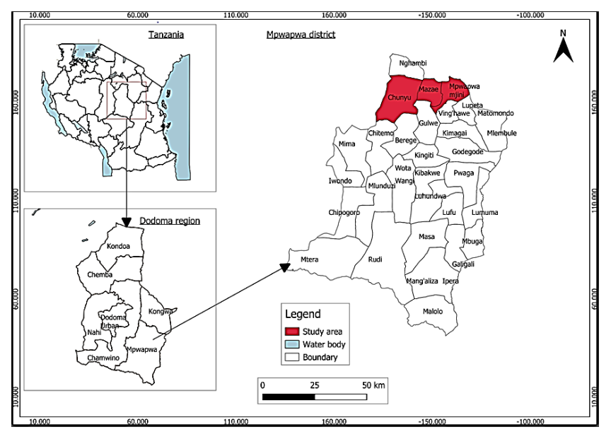
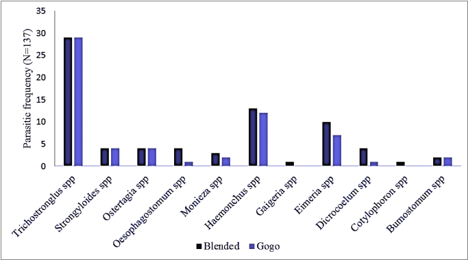
Gastrointestinal parasites (GIP) in goats pose a global challenge, resulting in significant negative impacts that affect health, productivity, and economic value. These parasites include nematodes, cestodes, and protozoa. The current study aimed to determine the prevalence and diversity of gastrointestinal parasites in goats found in Mpwapwa District. A cross-sectional study was carried out in Mpwapwa District from February to April 2025. A total of 200 faecal samples were obtained and analysed to detect gastrointestinal parasites. Additionally, a structured questionnaire was used to gather information from farmers on factors that might be linked to gastrointestinal parasitic infestations. The findings revealed a wide diversity of GIP in the study area, with a total of 11 GIP genera identified. The overall prevalence of GIP in goats was 61.5%. Breed-wise, Blended goats had a significantly (p<0.05) higher rate of gastrointestinal parasite infestation (71.3%) compared to Gogo goats (52.8%). Based on age, the prevalence was 65.6% in young animals and 59.6% in adults (p>0.05). Additionally, the physiological status of animals was significantly (p<0.05) associated with the prevalence of GIP. The body condition score did not influence parasitic prevalence (p>0.05). The most prevalent parasite was Trichostrongylus spp. At 42.3%, followed by Haemonchus and Eimeria spp., with prevalence of 18.2% and 12.4%, respectively. The current study revealed a high prevalence of parasites, indicating that GIP are a major challenge in goat production in the study area. Therefore, the application of proper control measures is recommended for the welfare of the goat production, but also addresses parasites that have zoonotic significance.
| Published in | Animal and Veterinary Sciences (Volume 13, Issue 5) |
| DOI | 10.11648/j.avs.20251305.15 |
| Page(s) | 152-161 |
| Creative Commons |
This is an Open Access article, distributed under the terms of the Creative Commons Attribution 4.0 International License (http://creativecommons.org/licenses/by/4.0/), which permits unrestricted use, distribution and reproduction in any medium or format, provided the original work is properly cited. |
| Copyright |
Copyright © The Author(s), 2025. Published by Science Publishing Group |
Gastrointestinal Parasite, Prevalence, Diversity, Goats
Variable | Category | Total number | Not Infected | Number infected | Prevalence (%) | X2 | P-value |
|---|---|---|---|---|---|---|---|
Breed | Blended | 94 | 27 | 67 | 71.3 | 7.14 | 0.007* |
Gogo | 106 | 50 | 56 | 52.8 | |||
Sex | Male | 26 | 14 | 12 | 46.2 | 2.972 | 0.085 |
Female | 174 | 63 | 111 | 63.8 | |||
Age | Adult | 136 | 55 | 81 | 59.6 | 0.411 | 0.44 |
Young | 64 | 22 | 42 | 65.6 | |||
Status | Weaner | 64 | 22 | 42 | 65.6 | 12.788 | 0.012* |
Dry does | 16 | 3 | 13 | 81.3 | |||
Breeding bucks | 6 | 5 | 1 | 16.7 | |||
Pregnant does | 82 | 29 | 53 | 64.6 | |||
Lactating does | 32 | 18 | 14 | 43.8 | |||
BCS | Fat | 43 | 14 | 29 | 67.4 | 3.534 | 0.171 |
Normal/good | 149 | 62 | 87 | 58.4 | |||
Poor | 8 | 1 | 7 | 87.5 | |||
Total | 200 | 77 | 123 | 61.5 | |||
Parasitic name | Frequency | Percent |
|---|---|---|
Trichostrongylus spp | 58 | 42.3 |
Haemonchus spp | 25 | 18.2 |
Eimeria spp | 17 | 12.4 |
Ostertagia/Teladorsagia spp | 8 | 5.8 |
Strongyloides spp | 8 | 5.8 |
Dicrocoelium spp | 5 | 3.6 |
Moniezia spp | 5 | 3.6 |
Oesophagostomum spp | 5 | 3.6 |
Bumostomum spp | 4 | 2.9 |
Cotylophoron spp | 1 | 0.7 |
Gaigeria spp | 1 | 0.7 |
Total | 137 | 100.0 |
GIP | Gastrointestinal Parasites |
GDP | Gross Domestic Product |
SEA | Small East Africa Breed |
BCS | Body Condition Score |
FEC | Faecal Egg Count |
OPG | Oocysts Per Gram |
EPG | Egg Per Gram |
| [1] | Ministry of Livestock and Fisheries, “The United Republic of Tanzania, Ministry of Livestock and Fisheries: Livestock Sector Transformation Plan (LSTP) 2022/23 - 2026/27,” 2022, p. 81. |
| [2] | K. Ngongolo and N. E. Mmbaga, “A study on the productivity and mortality rates of native and blended goats in Dodoma, Tanzania,” Pastoralism, vol. 12, no. 1, 2022, |
| [3] | S. Kusza, “A Review on Indigenous Goats of East Africa: A Case for Conservation and Management,” 2024. |
| [4] | A. Nguluma et al., “Typology and characteristics of indigenous goats and production systems in different agro-ecological zones of Tanzania,” Trop. Anim. Health Prod., vol. 54, no. 1, 2022, |
| [5] | R. Verma et al., “Epidemiology of Common Gastrointestinal Parasitic Infections in Goats reared in Semi-Arid Region of India,” no. February, pp. 39–45, 2018, |
| [6] | M. M. Gobena, “Production Performance, Challenges and Opportunity of Goat Production in Ethiopia,” vol. 50, pp. 26–35, 2016. |
| [7] | R. Day, A. Mohamed-brahmi, and F. Aribi, “Sustainable Goat Farming in Southeastern Tunisia : Challenges and Opportunities for Profitability,” pp. 1–25, 2025. |
| [8] | J. R. L. Mhoma, P. W. N. Kanyari, and J. M. Kagira, “The prevalence of gastro-intestinal parasites in goats in urban and peri-urban areas of Mwanza City, Tanzania,” vol. 12, no. December, pp. 191–196, 2011. |
| [9] | P. I. Zvinorova, T. E. Halimani, F. C. Muchadeyi, O. Matika, V. Riggio, and K. Dzama, “Prevalence and risk factors of gastrointestinal parasitic infections in goats in low-input low-output farming systems in Zimbabwe,” Small Rumin. Res., vol. 143, pp. 75–83, 2016, |
| [10] | W. Namutosi, J. Higenyi, E. Kizito, and M. Omodo, “Prevalence and Risk Factors of Gastrointestinal Parasite Infection in Goats in Sironko District, Eastern Uganda,” Uganda J. Agric. Sci., vol. 19, no. 1, pp. 1–14, 2020, |
| [11] | K. Shrivastava, A. P. Singh, K. Jadav, S. Shukla, and S. Prasad, “Caprine haemonchosis: optimism of breeding for disease resistance in developing countries,” 2022, |
| [12] | I. I. Abdul-rahman, P. I. Fuachie, and M. J. Tati, “Gastrointestinal Parasite Infection in Small Ruminants Relative to Host sex, Age and Husbandry System Under the Guinnea Savannah Vegatations,” vol. 38, no. 2, pp. 139–155, 2022. |
| [13] | T. J. Mpofu, “Gastrointestinal parasite infection intensity and hematological parameters in South African communal indigenous goats in relation to anemia,” vol. 13, pp. 2226–2233, 2020. |
| [14] | P. Admasu and L. Nurlign, “Prevalence of gastrointestinal parasites of small ruminants in Kuarit District, North West Ethiopia,” African J. Basic Appl. Sci., vol. 6, no. 5, pp. 125–130, 2014, |
| [15] | N. Moje, A. Gurmesa, and G. Regassa, “Gastro-intestinal Tract Nematodes of Small Ruminants : Prevalence and Their Identification in and Around Alage, Southern Ethiopia,” vol. 9, no. 3, pp. 65–72, 2021, |
| [16] | T. J. Mpofu, K. A. Nephawe, and B. Mtileni, “Prevalence of gastrointestinal parasites in communal goats from different agro-ecological zones of South Africa,” vol. 13, 2020. |
| [17] | L. J. M. Kusiluka, D. M. Kambarage, and L. J. S. Harrison, “Prevalence and seasonal patterns of coccidial infections in goats in two ecoclimatic areas in Morogoro, Tanzania,” vol. 30, pp. 85–91, 1998. |
| [18] | D. M. Kambarage, S. I. Kimera, L. J. M. Kusiluka, and M. M. A. Mtambo, “Prevalence of Eimeria and Cryptosporidium oocysts in cattle, sheep and goats in Morogoro Region, Tanzania,” vol. 2119, 2011, |
| [19] | D. Said et al., “Animal health constraints in dairy goats kept under smallholder farming systems in Kongwa and Mvomero Districts, Tanzania,” vol. 6, no. November, pp. 268–279, 2014, |
| [20] | G. S. Materu, J. S. Nzalawahe, M. E. Sengupta, and A. Stensgaard, “Prevalence, Distribution and Risk Factors for Trematode Infections in Domesticated Ruminants in the Lake and Southern Zones of Tanzania : A Cross-Sectional Study Prevalence, Distribution and Risk Factors for Trematode Infections in Domesticated Ruminant,” 2024, |
| [21] | H. Bedada, F. Gizaw, W. Negash, V. Medicine, and P. O. Box, “Preliminary Study on Small Ruminant GIT Helminthiasis in Select Arid and Semi-arid Pastoral and Agro-pastoral Areas of Afar Region, Ethiopia,” vol. 1, no. 1, pp. 1–9, 2018. |
| [22] | D. M. Komwihangilo, M. Jackson, Y. Munishi, B. S. A. Liheta, N. Livestock, and P. O. Box, “Situational analysis of smallholder goat production and marketing in Central Tanzania point towards the establishment of farmers ’ groups,” vol. 4, no. 12, pp. 356–364, 2012, |
| [23] | F. B. C. Njau, J. Lwelamira, and C. Hyandye, “Ruminant livestock production and quality of pastures in the communal grazing land of semi arid central Tanzania,” Livest. Res. Rural Dev., vol. 25, no. 8, pp. 1–13, 2016. |
| [24] | A. A.. Nor-Azlina and O. M., Sani, R. A. and Ariff, “Management Practices Affecting Helminthiasis in Goats,” vol. 34, no. November 2010, pp. 295–301, 2011. |
| [25] | D. K. Sharma, S. Paul, and K. Gururaj, “Gastrointestinal Helminthic Challenges in Sheep and Goats in Afro-Asian Region : A Review,” vol. 10, no. February, pp. 1–18, 2020, |
| [26] | H. Mruttu, C. Ndomba, G. Gebru, and J. Stapleton, “Animal breeding and genetics in the Tanzania livestock master plan,” 2015. |
| [27] | R. B. Onzima, R. Mukiibi, A. Ampaire, K. K. Benda, and E. Kanis, “Between-breed variations in resistance / resilience to gastrointestinal nematodes among indigenous goat breeds in Uganda,” pp. 1763–1769, 2017, |
| [28] | V. Philipo, A. M. Lupindu, and J. S. Nzalawahe, “Epidemiology and Molecular Identification of Coenurus cerebralis in Sheep and Goats in Mpwapwa District, Tanzania: Factors to Consider in Control Plans,” Vet. Med. Int., vol. 2024, no. 1, 2024, |
| [29] | I. Mengele, “Bovine brucellosis seroprevalence, farmers ’ awareness, practices and animal health extension services inputs in Mpwapwa district, Tanzania Bovine brucellosis seroprevalence, farmers ’ awareness, practices and animal health extension services inputs,” Tanzania Vet. J. · January 2023., no. January, 2023. |
| [30] | M. Thrusfield, veterinary epidemiology. 1995. Available: |
| [31] | M. Bhowmik et al., “Prevalence of Gastrointestinal Parasitic Infections in Sheep and Goats of Sandwip Island, Chattogram, Bangladesh,” Van Vet. J., vol. 31, no. 3, pp. 152–157, 2020, |
| [32] | J. Hansen and B. Perry, “The epidemiology, diagnosis and control of helminth parasites of ruminants,” Prev. Vet. Med., vol. 31, no. 1–2, pp. 161–162, 1994, |
| [33] | B. T. Paul et al., “veterinary sciences Risk Factors and Severity of Gastrointestinal Parasites in Selected Small Ruminants from Malaysia,” Vet. Sci. 2020, 7, 208, p. 14, 2020. |
| [34] | A. Rani, N. Begum, A. Alim, S. Malakar, T. Islam, and M. Zahangir, “Gastro-intestinal nematodes in goats in Bangladesh: A large- scale epidemiological study on the prevalence and risk factors,” Parasite Epidemiol. Control, vol. 9, p. e00146, 2020, |
| [35] | S. Islam, S. Hossain, A. R. Dey, A. Alim, S. Akter, and Z. Alam, “Epidemiology of gastrointestinal parasites of small ruminants in Mymensingh, Bangladesh,” vol. 7710, no. December, pp. 356–362, 2017. |
| [36] | A. Rahman, S. S. Labony, A. R. Dey, and M. Z. Alam, “An epidemiological investigation of gastrointestinal parasites of small ruminants in Tangail, Bangladesh,” vol. 15, no. 2, pp. 255–259, 2017, |
| [37] | M. A. Raza, M. Younas, and E. Schlecht, “Prevalence of gastrointestinal helminths in pastoral sheep and goat flocks in the cholistan desert of Pakistan,” J. Anim. Plant Sci., vol. 24, no. 1, pp. 127–134, 2014. |
| [38] | S. Mideksa, N. Mekonnen, Y. Muktar, V. Medicine, P. O. Box, and D. Dawa, “Prevalence and Burden of Nematode Parasites of Small Ruminants in and Around Haramaya University,” vol. 34, no. 5, pp. 644–651, 2016, |
| [39] | M. Adua, K. O. Idahor, and U. Sambo, “Role of physiological status of ruminant animals reared in Lafia metropolis on gastrointestinal parasites burden,” no. September, 2017, |
| [40] | N. M. F. Hassan, T. K. Farag, N. M. T. Abu El Ezz, and H. A. A. Abou-Zeina, “Prevalence assessment of gastrointestinal parasitic infections among goats in Giza Governorate, Egypt,” Bull. Natl. Res. Cent., vol. 43, no. 1, 2019, |
| [41] | K. Ashrafi, M. Sharifdini, Z. Heidari, B. Rahmati, and E. B. Kia, “Zoonotic transmission of Teladorsagia circumcincta and Trichostrongylus species in Guilan province, northern Iran: Molecular and morphological characterizations,” BMC Infect. Dis., vol. 20, no. 1, pp. 1–9, 2020, |
| [42] | D. K. Sharma, A. Mandal, B. Section, and E. Regional, “Factors affecting gastrointesti- nal parasite infections in goats in semi-arid rural ecosystems in India,” Vet. Sci. Dev. 2013, vol. 3, pp. 5–8, 2013, |
APA Style
Mgalla, I. C., Makingi, G., Nguluma, A. S., Mbaga, S. H. (2025). Prevalence and Diversity of Gastrointestinal Parasites in Native and Blended Goats from Mpwapwa District, Tanzania. Animal and Veterinary Sciences, 13(5), 152-161. https://doi.org/10.11648/j.avs.20251305.15
ACS Style
Mgalla, I. C.; Makingi, G.; Nguluma, A. S.; Mbaga, S. H. Prevalence and Diversity of Gastrointestinal Parasites in Native and Blended Goats from Mpwapwa District, Tanzania. Anim. Vet. Sci. 2025, 13(5), 152-161. doi: 10.11648/j.avs.20251305.15
AMA Style
Mgalla IC, Makingi G, Nguluma AS, Mbaga SH. Prevalence and Diversity of Gastrointestinal Parasites in Native and Blended Goats from Mpwapwa District, Tanzania. Anim Vet Sci. 2025;13(5):152-161. doi: 10.11648/j.avs.20251305.15
@article{10.11648/j.avs.20251305.15,
  author = {Imani Clemence Mgalla and George Makingi and Athumani Shabani Nguluma and Said Hemed Mbaga},
  title = {Prevalence and Diversity of Gastrointestinal Parasites in Native and Blended Goats from Mpwapwa District, Tanzania
},
  journal = {Animal and Veterinary Sciences},
  volume = {13},
  number = {5},
  pages = {152-161},
  doi = {10.11648/j.avs.20251305.15},
  url = {https://doi.org/10.11648/j.avs.20251305.15},
  eprint = {https://article.sciencepublishinggroup.com/pdf/10.11648.j.avs.20251305.15},
  abstract = {Gastrointestinal parasites (GIP) in goats pose a global challenge, resulting in significant negative impacts that affect health, productivity, and economic value. These parasites include nematodes, cestodes, and protozoa. The current study aimed to determine the prevalence and diversity of gastrointestinal parasites in goats found in Mpwapwa District. A cross-sectional study was carried out in Mpwapwa District from February to April 2025. A total of 200 faecal samples were obtained and analysed to detect gastrointestinal parasites. Additionally, a structured questionnaire was used to gather information from farmers on factors that might be linked to gastrointestinal parasitic infestations. The findings revealed a wide diversity of GIP in the study area, with a total of 11 GIP genera identified. The overall prevalence of GIP in goats was 61.5%. Breed-wise, Blended goats had a significantly (p0.05). Additionally, the physiological status of animals was significantly (p0.05). The most prevalent parasite was Trichostrongylus spp. At 42.3%, followed by Haemonchus and Eimeria spp., with prevalence of 18.2% and 12.4%, respectively. The current study revealed a high prevalence of parasites, indicating that GIP are a major challenge in goat production in the study area. Therefore, the application of proper control measures is recommended for the welfare of the goat production, but also addresses parasites that have zoonotic significance.
},
 year = {2025}
}
TY  - JOUR
T1  - Prevalence and Diversity of Gastrointestinal Parasites in Native and Blended Goats from Mpwapwa District, Tanzania

AU  - Imani Clemence Mgalla
AU  - George Makingi
AU  - Athumani Shabani Nguluma
AU  - Said Hemed Mbaga
Y1  - 2025/10/30
PY  - 2025
N1  - https://doi.org/10.11648/j.avs.20251305.15
DO  - 10.11648/j.avs.20251305.15
T2  - Animal and Veterinary Sciences
JF  - Animal and Veterinary Sciences
JO  - Animal and Veterinary Sciences
SP  - 152
EP  - 161
PB  - Science Publishing Group
SN  - 2328-5850
UR  - https://doi.org/10.11648/j.avs.20251305.15
AB  - Gastrointestinal parasites (GIP) in goats pose a global challenge, resulting in significant negative impacts that affect health, productivity, and economic value. These parasites include nematodes, cestodes, and protozoa. The current study aimed to determine the prevalence and diversity of gastrointestinal parasites in goats found in Mpwapwa District. A cross-sectional study was carried out in Mpwapwa District from February to April 2025. A total of 200 faecal samples were obtained and analysed to detect gastrointestinal parasites. Additionally, a structured questionnaire was used to gather information from farmers on factors that might be linked to gastrointestinal parasitic infestations. The findings revealed a wide diversity of GIP in the study area, with a total of 11 GIP genera identified. The overall prevalence of GIP in goats was 61.5%. Breed-wise, Blended goats had a significantly (p0.05). Additionally, the physiological status of animals was significantly (p0.05). The most prevalent parasite was Trichostrongylus spp. At 42.3%, followed by Haemonchus and Eimeria spp., with prevalence of 18.2% and 12.4%, respectively. The current study revealed a high prevalence of parasites, indicating that GIP are a major challenge in goat production in the study area. Therefore, the application of proper control measures is recommended for the welfare of the goat production, but also addresses parasites that have zoonotic significance.

VL  - 13
IS  - 5
ER  -
Department of Animal, Aquaculture and Range Sciences, Sokoine University of Agriculture, Morogoro, Tanzania
Department of Veterinary Medicine and Public Health, Sokoine University of Agriculture, Morogoro, Tanzania
Department of Animal, Aquaculture and Range Sciences, Sokoine University of Agriculture, Morogoro, Tanzania
Department of Animal, Aquaculture and Range Sciences, Sokoine University of Agriculture, Morogoro, Tanzania