Research Article | | Peer-Reviewed

Comparative Clinicopathologic Studies on Sheep Pox and Goat Pox in Bauchi and Gombe of North-Eastern Nigeria

Received: 28 January 2026     Accepted: 9 March 2026     Published: 16 April 2026
Views:       Downloads:
Abstract

In Nigeria, sheep pox and goat pox is a major problem. Sheep pox and goat pox diseases are highly contagious viral diseases of small ruminants caused by a virus of the Capripox genus. This study was aimed to comparatively evaluate the clinico-pathological changes in sheep and goats manifesting the clinical cases of the diseases. The design of the study was a cross sectional survey. A total of 166 sheep (66) and goats (100) were sampled purposively. Three millilitres of blood sampled was collected from the external jugular vein of affected and apparently healthy sheep and goats used for haematological analyses. Real-Time Polymerase Chain Reaction (RT-PCR) was used to confirm the presence of sheeppox and/or goatpox virus in 34 scrapped skin scrapings collected from sheep (14 samples) and goats (20 samples) that showed the characteristic of pock lesions. Dead sheep and goats were necropsied and fixed in 10% neutral-buffered formalin and were routinely processed for histopathology. Clinical signs observed were fever (high rectal temperatures of 40 to 41.5°C in sheep and 38 to 40°C in goats), papules, and abortion in goats among others. The mean PCV value of sheep and goats with pock lesions (24.07±5.36%) and (27.63±5.24%), respectively were significantly lower than those of apparently healthy sheep (33.09±4.24%) and goats (31.66±2.7%). The mean HBC value of sheep and with pock lesions (8.80±2.49g/dl) was significantly lower than those of apparently sheep (11.30±1.62g/dl), while there was no significant difference between the HBC of goats with pock lesions (12.78±2.81g/dl) and apparently healthy goats (11.91±1.14g/dl). The mean RBC of sheep value (6.46±2.55×10¹²/L) was significantly lower than that of apparently healthy sheep (9.06±2.24 x 10¹²/L), The mean values for WBC count, absolute neutrophils and lymphocyte were also affected. Of the 34 skin samples collected and analyzed for the presence of sheep/goat pox viruses, 30 (88.2%) yielded positive using universal Capripox primers. Of the 30 positive samples, 21 were further subjected to sheep pox specific primers, 18 (85.7%) positive were obtained for sheeppox specific primers and no sample yielded positive result to goat specific primers. Overall, 88.2% of sheep and goat populations showing pock lesions were confirmed for sheeppox. The results of this study suggest that sheeppox virus was responsible for the disease outbreaks in sheep and goats from the study areas. Sheep pox disease has the potential of causing significant alteration in the hematological parameters resulting in anemia and immunosuppression. Yearly vaccination of sheep and goats is therefore recommended.

Published in Animal and Veterinary Sciences (Volume 14, Issue 2)
DOI 10.11648/j.avs.20261402.11
Page(s) 25-35
Creative Commons

This is an Open Access article, distributed under the terms of the Creative Commons Attribution 4.0 International License (http://creativecommons.org/licenses/by/4.0/), which permits unrestricted use, distribution and reproduction in any medium or format, provided the original work is properly cited.

Copyright

Copyright © The Author(s), 2026. Published by Science Publishing Group

Keywords

Sheep, Goat, Pox, Virus, Bauchi, Gombe, North-East, Capripox, Immunosuppression

1. Introduction
1.1. Background of the Study
Sheep pox and goat pox are highly contagious viral diseases caused by a virus of the genus Capripoxvirus, family Poxviridae, and can infect sheep, goats, and other domestic and wild ruminants . It is a World Organization for Animal Health (WOAH), formally Office International des Epizooties (OIE), list A disease . Sheep pox and goat pox are prevalent in many regions of Asia, Africa, and the Middle East, where they pose a significant threat to small ruminants’ production and trade . The diseases can cause reduction in milk production, impaired reproductive performance and mortality in infected sheep and goats , leading to severe economic losses for farmers and communities. Poxvirus infections have ability to reduce trade, export, and development of intensive livestock production. It also prevents the import of new breeds . The host preferences of sheeppoxvirus (SPV) and goatpoxvirus (GPV) can vary among different isolates, with some causing disease exclusively in sheep or goats and others affecting both species . In Nigeria, sheep and goat pox (SGP) has been reported with prevalence rates of 34% and case fatality rates of 53% respectively . Though the diseases had been reported, but they are under documented.
Clinical manifestations of sheeppox or goatpox infection include fever, respiratory distress, and the development of the characteristic pock lesions in the skin, mucous membranes, and internal organs . In severe cases, the diseases can lead to death in affected animals . The infections are mild and common in indigenous breeds, although symptoms may be more severe in lambs and kids, stressed animals, animals with concurrent infections or naïve flock . Morbidity and mortality vary with the species and breed of the animal, its immune status to Capripoxviruses, and the strain of the viruses. Mortality may be up to 50% in a fully susceptible flock and as high as 100% in young animals .
The diagnosis of sheep pox and goat pox can be challenging without laboratory tests, even though they are from the same genus but they are distinct from each other and can cause cross infection between species. Both diseases share many clinical and pathological features together . Diagnosis involves clinical examination, lesion observation, histopathology, and laboratory confirmation using techniques such as polymerase chain reaction (PCR) and virus isolation .
Prevention and control of sheep pox and goat pox rely mainly on vaccination, strict biosecurity measures, and early detection and isolation of infected animals . Several commercial vaccines are available for the prevention of sheep pox and goat pox, and they have been shown to provide effective protection against the diseases . Unfortunately, vaccination of sheep and goats with sheep pox and goat pox vaccines is not commonly practiced by farmers in Nigeria in spite of frequent outbreaks of the diseases in many parts of the country. Previous studies reported a prevalence of 34% and case mortality rate of 54% in sheep and goats within Nigeria but report in Punjab, Pakistan revealed morbidity of 75-100% and case fatality of 10-85% depending on the virulence of the virus .
1.2. Statement of the Research Problem
Sheeppox and goatpox are considered to be emerging and significant viral diseases that affect small ruminants and cause substantial economic losses in small-holder farmers in Nigeria. However, there has not been a comprehensive study on the comparative clinico-pathological changes in sheep pox and goat pox diseases in Nigeria. Most veterinary clinicians and farmers presume that the diseases have the same pattern and magnitude of spread in both animal species. This knowledge gap hinders the development of effective control and prevention strategies tailored to these diseases in the country.
1.3. Aim of the Study
The aim of the study is to comparatively evaluate the clinicopathological changes in sheep pox and goat pox infected sheep and goats in Bauchi and Gombe States of northern Nigeria.
1.4. Specific Objectives
The specific objectives of this study are to;
1) Establish the diagnosis of suspected sheep pox and goat pox using clinical observations and Real-Time PCR to confirm the diseases in the study areas.
2) Compare the gross and histopathological changes observed in sheep and goats infected with poxviruses in the study areas.
3) Compare the hematological changes in sheep and goats with poxvirus diseases in the study areas.
2. Materials and Methods
2.1. Study Location
The study was carried out in Bauchi and Gombe States of North-East, Nigeria located on Longitude 10.3060°N, Latitude 9.8404°E and Longitude 10.3638°N, Latitude 11.1928°E, respectively.
2.2. Ethical Approval
Approval for the use of animal for this research was obtained from the National Veterinary Research Institute, Vom-Institutional Animal Care and Use Committee (NVRI-IACUC) with Approval Reference Number: NVRI-IACUC-AEC/02/155/24.
2.3. Materials
Animals
Sheep and goats of both sexes and of different breeds and ages.
2.4. Methods
Study Design
The design of the study was cross sectional survey of sheep and goats of both sexes and of different breeds and ages manifesting lesions suggestive of sheeppox and goatpox (SGP) which were purposively sampled from households and farms during the period of the disease outbreaks. A minimum of 166 sheep and goats were sampled, as calculated using Raosoft™ Sample Size Calculator (www.roasoft.com), based on a previous study on SGP diseases in Bauchi State, where 2.9% sero-prevalence rate was reported , also assuming a 2.9% prevalence rate for Gombe State.
2.5. Examination of Animal
The animals were examined for lesions manifested over their bodies suggestive of sheeppox and goatpox, and were scored according to severity as follows: mild (+) when they were sparsely distributed over the body, moderate (++) and severe (+++) when the whole body is involved. Rectal temperature (degree Celcius) was taken before sample collection.
2.6. Sample Collection
2.6.1. Blood Sample Collection
Exactly three milliliters (3ml) of blood sample for complete hematology was aseptically collected from the jugular vein of sheep and goats into an EDTA bottles; with 7 Samples each collected from apparently healthy sheep and goats to serve as control whose haematological parameters were compared with those of the sick animals. The samples were placed in ice-packs and transported to the National Veterinary Research Institute, Vom for hematological analysis were the Packed cell volume (PCV) was determined by the microhaematocrit method , Red Blood Count (RBC) counts were obtained by haemocytometer method , White Blood Cell (WBC) counts were obtained by haemocytometer method , Haemoglobin Concentration (HBC) was measured by cyanomethaemoglobin method and Differential Leucocyte Count (DLC) was done using Leishman’s technique .
2.6.2. Skin Scrapings/Scabs Collection
Sterile surgical bled were used for collection of skin scrapings/scabs into a plain vacoutainer tubes and were transported in cool chain, to minimize viral deactivation by high environmental temperatures, to the NVRI Viral Research Laboratories for Virus Identification using Real Time Polymerase Chain Reaction (RT-PCR) method of analysis as described by .
2.6.3. Tissue Processing for Histopathology Examination
Post mortem examinations were conducted on the sheep and goats that died from the suspected diseases. After gross examination of organs, the following tissues, liver, lung, kidney, spleen, skin and the fore stomach with intestine, were harvested and placed in 10% neutral buffered formalin containing containers for further processing at the Histopathology Laboratory Unit of Veterinary Pathology Department, NVRI. At the histopathology lab, the samples were trimmed and placed in labeled cassettes appropriately for dehydration. The tissue specimens were immersed in series of ethanol solutions of increasing grade/concentration (from 60-100%) until pure, water-free (absolute) ethanol was reached for specific of time (15 minutes in each ethanol concentration). Tissue specimens were removed from ethanol and immersed in xylene. Tissues were immersed for 20 minutes each in three different jars of xylene. The tissues were further impregnated with paraffin wax (embedding agent) in a beaker placed in a hot air oven for 6-8 hours. After tissues had been dehydrated, cleared and infiltrating with embedding material, they were now placed in leuckhart molds/blocks for cooling. The paraffin blocks were sectioned using rotating thermofisher microtome (USA) with 4-5 micro-meter thickness. Sectioned tissues were place in warm water bath and picked up from the water bath and placed on a clean grease free glass slide in hot air oven for 15 minutes to enable the sections adhere properly on the slide. Slides were labeled appropriately with non-removable ink. Slides were cleared by dipping ten times in xylol to remove wax and rinsed in graded concentrations of alcohol (65-95%) to remove xylol before staining. The slides were stained with Hemtoxylin and eosin stain and allowed to dry. The stained sections on the glass slides were covered with a thin piece of cover slip. The slides were allowed to dry for 15 minutes in a hot air oven. The slides were examined under a microscope at different fields and pictures were captured.
2.7. Molecular Detection of SPV and GPV
Tissue sample from skin scrapings/scabs were homogenized using sterile glass sand and phosphate buffered saline (PBS) to enhanced virus DNA extraction easily from the skin scabs, the homogenates were placed into sterile Bijou bottle centrifuge at 2000 rpm for 5 minutes, the supernatant was removed using needle and syringe and were transferred into a sterile cryovial and stored in -20°C. Virus Deoxyribonucleic acid (DNA) extraction was done by the QIAamp DNA minikit protocol for DNA extraction following the steps described in QIAGEN kit handbook. DNA amplification was set in Masters Mix and was votex using Stuart designed in the United Kingdom (UK) and were placed in the PikoReal 24 Thermo Scientific PCR machine with system made in United States of America (USA) and assembled in Germany at a required cycle, time and temperature compared with positive and negative samples for comparison. Samples were allowed to run for 45 cycles at the following temperatures and times; 95°C for 3mins, 95°C for 15mins, 60°C for 15mins and 72°C for 15mins and the following primers and sequences were used for both forward and backward reactions.
Capri-F AAAACGGTATATGGAATAGAGTTGGA,
Capri-R AAATGAAACCAATGGATGGGATA,
SPPV-F AGGTACAAAATAATACCAACBATTC,
SPPV-R GTTGATTTTTCAACATTTATGTAG,
GTPV-F CATTTGTTGATATAAACG,
GTPV-R CTARAGATTTAGAAACRACGGTAAAA
(SigmaAldrich.com/oligolearning, 2023).
The virus identification was done using PikoReal 24 Thermo scientific (Germany) RT-PCR method as described and validated . RT-PCR methods for direct capripox genotyping without the need for gene sequencing as described Positive samples were further subjected to specific primers of sheep pox and goat pox to differentiate between the virus species .
2.8. Data Analysis
Data generated from the study were subjected to Chi-Square to check the association between the lesions and the positive samples (PCR positive), Student t-test was used to compare hematological parameters between healthy and sick animals and data were analyzed by descriptive statistics. Significance was accepted at p < 0.05. Summary of results was presented as means with standard deviation of mean, tables and bar charts. The data was analyzed using Statistical Package for Social Sciences (SPSS) version 16.0.
3. Results
3.1. Clinical Signs
The observed clinical signs include very high body temperatures of 40 to 41.5°C in sheep and 38 to 40°C in goats, loss of appetite, emaciation, lethargy, dyspnea, mucopurulent nasal and ocular discharges, dry scabs on healing wounds, abortion in goats (particularly noticed in Bauchi State), and mortalities (Table 1). The pock lesions measured from 1 to 4cm in diameter and appeared mostly as small and big coalesced nodules distributed all over the body of the severely affected sheep and goats (Figures 1 and 2). Nodules erode forming ulcers and necrotic scabs containing dry pus underneath.
Specifically, in sheep, the pock lesions were found on the face, dorsal ear, maxilla and mandibular regions and on the oral commissures (Table 2). Other gross lesions include enlarged/swollen lymph nodes, engorged gallbladder, pale kidneys with areas of congestion, pale and enlarged spleen; bilateral flat gray nodules sparsely distributed on the lungs, and clotted blood in the trachea with flabby myocardial musculature (Table 3). The lesions seen in goat were crusts formation and scabs which sloughed off easily, frothy exudates from the upper respiratory track (larynx and pharynx) down to the entire airway segments, bilateral congestion of the lungs, the intestinal mucosae were severely haemorrhagic, the livers were swollen and congested, engorged gallbladder, enlarged and congested spleen, swollen lymph nodes, flabby cardiac musculature and hyperemic areas in the rumen and reticulum (Table 3).
Table 1. Degree (%) of manifestation amongst each species.

Clinical signs

Sheep

Goats

Rectal temperature

40-41.5°C

38-40°C

In-appetence

Present

Present

Emaciation

Present

Present

Lethargic

Present

Present

Difficulty in breathing

Present

Present

Ocular discharges

Present

Present

Formation of crust

Present

Present

Dry scabs on healing wounds

Present

Present

Ulceration

Present

Absent

Alopecia

Present

Present

Abortion

Absent

Present

Table 2. Distribution of pock lesions on body parts of poxvirus-infected sheep and goats.

Body location

Sheep

Goat

Face

++

++

Ears

+

++

Mouth (oral commissure)

++

+++

Nose

++

+++

Neck

+

++

Mandible

++

++

Limbs

++

++

Back (dorsum)

++

++

Tail

+

++

Perineum

+

++

Prepuce/testes

++

+++

Udder

++

+++

Ventral abdomen

+

++

Lateral abdomen

++

++

Inner thigh

++

++

Lateral thigh

++

++

Key: + = mild, ++ = moderate and +++ = severe
Table 3. Degree of occurrence of the gross lesions observed in poxvirus-infected sheep and goats.

Body location/organ

Sheep

Goat

Skin

+

++

Nasal block with thick mucoid exudates

++

++

Swollen lymphnode

++

++

Engorged gallbladder

+

-

Pale and enlarged spleen with areas of congestion

++

++

Congestion of the lungs

+

++

Ocular discharges

-

+

Congested and swollen liver

+

++

Hyperaemia of the rumen and reticulum

-

+

Frothy exudates in the entire trachea down to the alveoli

-

++

Congested areas of the intestinal mucosae

-

+++

Flabby cardiac musculature

+

++

Congested kidney

-

++

Hyperaemic areas on the gum

-

+

Key: +++ Severe, +++ Moderate, + Slight, - Nil
3.2. Hematological Parameters
In sheep, the mean values for PCV and haemoglobin concentration were significantly lower (p ˂ 0.05) in the sheep with the disease compared to the values of the healthy sheep. Similarly, the mean values for red blood cell (RBC) count and total white blood cell (WBC) count were significantly lower (p ˂ 0.05) in the sheep with sheeppox compared to the values in healthy sheep. Furthermore, the mean values for absolute neutrophil and lymphocyte counts were significantly lower (p ˂ 0.05) in the sheep with sheeppox compared to the values in healthy sheep (Table 4).
In goats, the mean PCV value was significantly lower (p ˂ 0.05) in the goats with goatpox compared to the values in healthy goats. There were no significant differences (p ˃ 0.05) in the mean values for haemoglobin concentration and RBC count between the healthy and the goats with goatpox. However, the mean value for WBC count of the goats with goatpox was significantly lower (p ˂ 0.05) than that of the healthy goats. The mean values for absolute neutrophil and lymphocyte counts of goats with goatpox were significantly lower (p ˂ 0.05) than those of the healthy goats (Table 5).
Table 4. Hematological parameters of healthy sheep and those with lesions of poxvirus infection.

Parameters

Mean±SD

Pvalue (2-tailed)

Healthy

Poxvirus infected sheep

PCV (%)

33.09±4.24

24.07±5.36

0.001

HbC (g/dL)

11.30±1.62

8.80±2.49

0.020

RBC Count (×1012/L)

9.06±2.24

6.46±2.55

0.030

MCV (FL)

37.4

37.3

0.00

MCHC (g/dL)

34.1

36.6

0.00

MCH (pg)

12.5

13.2

0.00

Total WBC Count (×109/L)

15.76±5.74

5.72±4.20

0.002

Neutrophils Count (×109/L)

7.62±2.99

3.14±3.01

0.007

Lymphocytes Count (×109/L)

8.27±3.74

2.72±1.78

0.007

Table 5. Hematological parameters of healthy goats and those with lesions of poxvirus infection.

Parameters

Mean±SD

P value (2-tailed)

Healthy

Poxvirus infected goats

PCV (%)

31.66±2.70

27.62±5.24

0.019

HbC (g/dL)

11.91±1.14

12.78±2.81

0.274

RBC Count (×1012/L)

6.22±1.85

5.63±1.53

0.469

MCV (FL)

50.9

49.1

0.00

MCHC (g/dL)

35.4

46.3

0.00

MCH (pg)

19.1

22.7

0.00

Total WBC Count (×109/L)

15.65±3.92

7.15±3.28

0.001

Neutrophils Count (×109/)

6.61±3.10

3.26±1.64

0.029

Lymphocytes Count (×109/)

8.84±2.88

3.82±2.35

0.020

3.3. Real-Time Polymerase Chain Reaction
A total of 34 samples (skin scrapings) were collected from the sheep and goats. Out of the 34 samples, 30 were positive and 4 were negative to poxviruses using the General Capripox Primers. Out of the 30 positive samples, 18 were from goats while 12 were from sheep. (Table 6).
Table 6. RT-qPCR detection of poxvirusus in general capripox primers.

Animal specie

Bauchi

Gombe

Total

Sheep

5 (14.7%)

7 (20.6%)

12 (35.3%)

Goat

8 (23.5%)

10 (29.4%)

18 (52.9%)

Total

13 (38.2%)

17 (50%)

30 (88.2%)

Twenty-one (21) of the positive samples further subjected to sheep pox specific primers yielded 18 positives and 3 negatives. The same numbers of samples were equally subjected to goat pox specific primers and none was positive.
There was an association between the pock lesions and the presence of the pox virus in the sheep and goats. There was a positive/direct relationship between pox virus infection and the packed cells volume, haemoglobin concentration, red blood cell count, total white blood cell counts and absolute neutrophil count. An indirect relationship was observed between poxvirus infection and absolute lymphocyte count.
3.4. Gross Lesions Histopathological Changes
The gross lesions observed in affected sheep were, engorged gallbladder, bilateral flat gray nodules sparsely distributed on the lungs, enlarged/swollen lymph nodes, pale kidneys with areas of congestion, pale and enlarged spleen. The gross lesions were more apparent on the skin and in the kidneys and lungs (Figure 1).
The gross lesions observed in the goats were frothy exudates from the upper respiratory track (larynx and pharynx) down to the entire airway segments, bilateral congestion of the lungs, the intestinal mucosae were severely haemorrhagic, the livers were swollen and congested, engorged gallbladder, enlarged and congested spleen, swollen lymph nodes, flabby cardiac musculature. The gross lesions were more pronounced on the skin and in the heart, intestines, lungs, kidneys and spleen (Figure 2).
In sheep showing pox lesions, sub-epidermal microabscesses were observed along with degeneration of the spinosum cells in the epidermis, furunculosis and hyperkeratinization. Cavities (oedematous areas) were observed in the dermis (Figure 3). The skin lesions in goats comprised degeneration and desquamation of keratinocytes in the epidermis, presence of papules in the epidermis and dermis as well as hyperaemia and oedema of the dermis (Figure 3). The heart tissue of affected sheep had severe degeneration and coagulative necrosis of the myocytes. Similar lesions were observed in the goats with the pock lesions. However, the myocardial necrosis in the goats was more extensive compared to that of the sheep (Figure 4). The small intestine of the affected sheep showed no obvious changes, microscopically. However, the duodenum of the affected goats was characterized by necrosis and atrophy of the villi (Figure 5). Moderate renal tubular degeneration and necrosis were observed in sheep with pock lesions, but in the goats with the disease, the kidney lesions were more severe and consisted of severe congestion of the cortex, tubular necrosis, and dilation of Bowman’s space (Figure 6). In liver of the affected sheep, there was moderate coagulative necrosis of centrilobular and periportal hepatocytes. Whereas severe centrilobular vacuolar degeneration with congestion of the central vein, severe sinusoidal dilation and congestion of the portal vein were observed in the goats (Figure 7). There were no lesions in the spleen of the affected sheep, microscopically, but severe lymphoid depletion, congestion and oedema were observed in the spleen of affected goats (Figure 8). There was thickening of the interstitial septa of the alveoli, presence of exudates in the lumen of the bronchioles and the alveoli with massive infiltration of mononuclear inflammatory cells in the septa and the lumen of sheep lungs. There were thickening of the interalveolar connective tissues and infiltration of the connective tissues with mononuclear inflammatory cells observed in the lungs of goats (Figure 9).
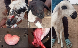
Figure 1. Some of the gross lesions observed in sheep infected with poxvirus. A-B: Alopecia, erosions, ulcerations and scar tissue formation on the face with mucopurulent nasal discharge. C: nodules underneath the skin of the face, dorsal part of the ear, neck and forelimbs. D: pale kidney. E: consolidated lungs (stage 3 consolidation or gray hepatization) with rib imprints on the costal surface.
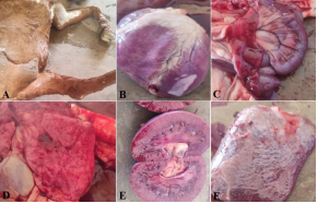
Figure 2. Some of the gross lesions observed in goats infected with poxvirus. A: nodules underneath the skin of the udder, ventral abdomen and limbs. B: patchy pale areas in the epicardium. C: Severe haemorrhage in the duodenum. D: Severely hyperaemic lung lobules. E: Kidney showing congestion of the medulla. F: Enlarged dome-shaped and mottled spleen.
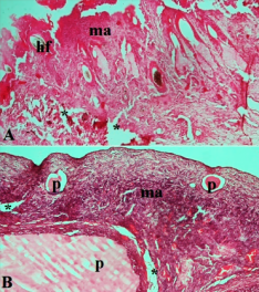
Figure 3. Skin section of sheep (A) and goat (B) infected with sheep poxvirus. Note the epidermal necrosis and sub-epidermal micro-abscesses containing predominantly mononuclear inflammatory cells, papules (p) containing hemorrhagic fluid in goat with dermal edema (asterisks), hf = hair follicle. H&E stain ×100.
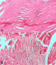
Figure 4. Heart tissue sections of sheep (A) and goat (B) infected with sheep poxvirus. Note extensive necrosis of the myofibres in (A) and moderate necrosis of the myofibres in (B). H&E stain ×200.
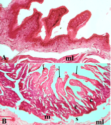
Figure 5. Intestinal sections of sheep (A) and goat (B) infected sheep poxvirus. Note severe necrosis of villi in the duodenum of goat (arrows). Ml = muscular layer, s = submucosa, m = muscularis mucosa, c = crypts. H&E stain ×100.
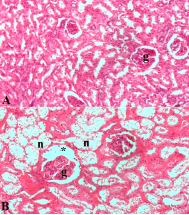
Figure 6. Kidney tissues of sheep and goat infected with sheep poxvirus. Note the severe tubular necrosis (n), atrophy of a glomerular tuft and dilation of the Bowman’s space (black asterisk), congested cortex with edema (white asterisk) in the goat, g = glomerulus. H&E stain ×200.
Figure 7. Liver sections of sheep (A) and goat (B) infected with sheep poxvirus. Note moderate diffuse hepatocellular necrosis in sheep and severe centrilobular vacuolar degeneration with congestion of the central vein (cv), severe sinusoidal dilation (s) and congestion of the portal vein (pv) in the goat. H&E stain ×200.
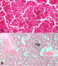
Figure 8. Splenic tissue sections of sheep (A) and goat (B) infected with sheep poxvirus. A: severe haemosiderosis in the red pulp B: severe congestion of the spleen with lymphoid depletion in the white pulp (wp). H&E stain, (A = ×400, B = ×100).
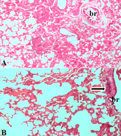
Figure 9. Lungs tissues of sheep (A) and goat (B) infected with sheep poxvirus, note severe interstitial pneumonia with formation of non-encapsulated micro-granulomas in sheep (encircled areas) composed of lymphocytes, macrophages and plasma cells. Note the peri-bronchiolar infiltrates in the goat (arrow), br = bronchiole H&E stain ×100.
4. Discussion, Conclusion and Recommendations
4.1. Discussion
In this study, theclinical signs such as high rectal temperature, loss of appetite, emaciation, lethargy, difficulty in breathing, ocular discharges, formation of crust around the mouth, skin ulcerations, alopecia and abortion observed are in agreement withclinical signs in diseases caused by viruses of the capripox genus, as reported by in Algeria; in India; and in Egypt. In the present study, lesions were found distributed all over the body in the severely infected animals and contrast with the report of who reported lesions on hairless areas of the body in Northern Vietnam. In the present study, high rectal temperature (pyrexia) of between 40 to 41.5°C reported in sheep and between 38 to 40°C in goats which is a common finding in capripox infection were comparable with the study done by in sheep and goats in Jammu Region of India, where high rectal temperature (pyrexia) of 41.5°C and 40°C respectively was reported.
In the present study, pock nodules surrounded by a clear zone of hyperaemia with some coalesced to form a lymphoma-like lesions were similar to those described by . Also the discrete flat grey nodules observed in the lungs of sheep in this study were similar to findings reported previously this because lungs is the most affected organ in capripox infections .
The occurrence of abortion in goats further underscores the economic importance of poxvirus disease in goats as well as the relative severity of the disease in goats compared to sheep. This finding is in line with previous study in Northern Vietnam where the lesions of poxvirus disease were observed to be more pronounced in goats than in sheep . Similarly, all the gross lesions observed on the body and other internal organs in the present study were similar to those previously reported in experimental animals in Jammu and this may suggest that both naturally and experimentally-infected animals may manifest similar poxvirus lesions, in terms of nature of lesions, its distribution and severity. The findings of this study also suggest that the disease has a significant potential to infect sheep and goats of different species, ages and sexes, as observed during the field survey, with highly devastating impact on young animals due to their weak immune status. These findings were similar to the reports of and , in which morbidity of 90% and mortality of 50% were in sheep and goats respectively.
Although there are no reports on the hematological changes associated with sheep and goat pox virus infections in Nigeria, to the best of my knowledge, in the present study, the PVC, RBC and WBC count of infected sheep/goats were significantly lower than apparently healthy sheep and goats. The cause of the decrease in the hematological parameters in the affected sheep and goats can be associated by a combination of the primary virus infection and secondary complications with bacterial infection like Pasteurella spp or enteritis leading to toxemia, which suppresses bone marrow activity and promotes the destruction of RBCs. Concurrent parasitic disease with Haemonchus contortus is likely to influence blood loss in the already stressed affected animals with Capripox infection. However, hemoglobin concentration was not affected in the poxvirus-infected sheep and goats.
The visceral organs of sheep in this present study were seen to have generalized paleness with whitish nodules sparsely distributed on the lungs while those in goats were marked with severe congestion similar to those found by .
The histopathological lesions observed in the liver in the present study, although were non-specific, but they are similar to the typical lesions of capripox infection reported in previous studies . There was thickening of the interalveolar septae in the lung of goats which were also described in previous reports .
Histological sections of the kidney in this study revealed degeneration and necrosis of renal tubules and interstitial nephritis and these kidney lesions were similar to those observed in sheep and goats in previous studies . It was postulated that an inflammatory response secondary to viremia and involvement of the kidney tubules lead to a pronounced interstitial reaction . Tatrophy of the glomerular tuft in goats in the present study was also earlier reported by and .
Skin scrapings/scabs from sheep and goats manifesting typical signs and lesions of the diseases were collected for RT-PCR diagnosis because detection of the viral presence is possible on dry scabs. The high percentage of positive cases 30 (88.2%) indicates the high sensitivity of capripox primers, and this is comparable to report of in Northern Vietnam, who detected a positive prevalence of 79.6% using PCR but different from work done in three states of Northern Nigeria by who detected a sero-positive prevalence of only 2%. This was because complement ELISA was deployed and serum rather than skin scabs/scraping from animals were used instead of PCR which have been chosen to be the best method for the detection for pox viruses for its sensitivity and specificity. Out of the 21 samples from both sheep and subjected to sheeppox-specific primers, 18 (85.7%) samples were positive. This confirms that sheeppox virus causes the disease in both sheep and goats. Furthermore, no sample yielded positive when they were subjected to goatpox-specific primers. This suggests that sheep poxvirus was responsible for the outbreak of poxvirus diseases in both sheep and goats in Bauchi and Gombe States.
4.2. Conclusion
The findings in this study have shown that infection with Sheeppox virus was responsible for the diseases observed in both sheep and goats in the study areas. The results obtained from this study further demonstrated the capacity of the infection with sheeppox virus to cause significant changes in the hematological parameters with possible consequences of anemia and immunosuppression. The PCR-positive samples obtained from the study areas confirmed that sheep poxvirus disease is endemic in the study areas. Although the histopathological changes in the skin and visceral organs are non-specific, their occurrence may modify the pathogenesis and influence the prognosis of the disease in sheep and goats.
4.3. Recommendation
Farmers should be encouraged to always vaccinate their animals against sheep poxvirus disease (which also protects against goat poxvirus disease) because vaccination is an effective means of disease prevention and control. More extensive research is necessary to further elucidate both the prevalence and pathogenesis of the diseases in sheep and goats in Nigeria.
4.4. Contributions to Knowledge
1) Sheeppoxvirus is mostly responsible for the outbreak of sheeppoxand goatpox diseases in Bauchi and Gombe States of Northern Nigeria.
2) Sheeppoxvirus infection causes significant alteration in the hematological parameters of infected animals.
Abbreviations

WOAH

World Organization for Animal Health

SPV

Sheep Pox Virus

GPV

Goat Pox Virus

PCR

Polymerase Chain Reaction

OIE

International des Epizooties

NVRI

National Veterinary Research Institute

SGP

Sheep and Goat Pox

EDTA

Ethylene Diamine Tetraaceticacid

PVC

Park Cell Volume

RBC

Red Blood Count

WBC

White Blood Count

DLC

Differential Leucocyte Count

HBC

Haemoglobin Concentration

Acknowledgments
The authors are eternally grateful to the farmers who willingly agreed for their animals to be sampled during the disease outbreak and as well as provided us with the necessary information required during our field trips.
Author Contributions
Logyang Lot Emmanuel: Conceptualization, Data curation, Investigation, Writing – original draft
Anyogu Davinson Chuka: Supervision, Methodology, Writing – review & editing
Adeyinka Adedeji: Methodology, Validation
Arinzechukwu Stephen Ezema: Data curation, Formal analysis
Mafulul Joshua Bako: Validation
Bolajoko Mohammed Bashiru: Methodology
Shuaibu Rabiata Haruna: Resources
Idoko Felix: Validation
Choji Tobias Peter Pwajok: Visualization
Maguda Adrian: Validation
Moses Gyang Davou: Formal Analysis
Chah Kennedy: Supervision, Methodology, Writing – review & editing
Shoyinka Vincent: Conceptualization, Methodology
Data Availability Statement
Relevant data and information about the present study will be provided on request.
Conflicts of Interest
The authors declare no competing interests.
References
[1] Abd-Elfatah, E. B., El-Mekkawi, M. F., Bastawecy, I. M., &Fawzi, E. M. (2018). Identification and phylogenetic analysis of sheep pox during an outbreak of sheep in Sharkia Governorate, Egypt. Genetics and Molecular Research, 17(2).
[2] Abdel-Ghaffar, K. H. S., &Hassanein, K. M. A. (2009, October). Pathological study of malignant form of sheep pox. In 10th Scientific Congress of Cattle Diseases. Faculty of Veterinary Medicine, Assiut University, Assiut, Egypt.
[3] Abubakar, M., Mahapatra, M., Muniraju, M., Arshed, M. J., Khan, E. H., Banyard, A. C., &Parida, S. (2015). Peste des petitsruminants virus, Asia and Africa. Emerging Infectious Diseases, 21(2), 202–208.
[4] Achour, H. A., Bonguedour, R., Bouhbal, A., &Guechtouli, A. (2000). Comparative study of immunizing ability of some attenuated strains of sheeppox virus and sensitizing vaccine. Revue Scientifiqueet Technique (International Office of Epizootics), 19(3), 773–783.
[5] Adedeji, A. J., Moller, J., Meseko, C. A., Adole, J. A., Tekki, I. S., Shamaki, D., & Hoffmann, B. (2019). Molecular characterization of capripoxviruses obtained from field outbreaks in Nigeria between 2000 and 2016. Transboundary and Emerging Diseases, 66(3), 1629–1641.
[6] Aswini, B. S. (2015). Pathological and molecular diagnosis of sheep pox (Master’s thesis). Sri Venkateswara Veterinary University, Tirupati, Andhra Pradesh, India.
[7] Babiuk, S., Bowden, T. R., Boyle, D. B., Wallace, D. B., &Kitching, R. P. (2008). Capripoxviruses: An emerging worldwide threat to sheep, goats, and cattle. Transboundary and Emerging Diseases, 55(7), 263–272.
[8] Balamurugan, V., Krishnamoorthy, P., Raju, D. S., Kumanan, K., Raj, G. D., Nachimuthu, K. & Singh, R. K. (2014). Capripox: An emerging and re-emerging zoonosis in India. VirusDisease, 25(4), 398–404.
[9] Dar, L. M., Darzi, M. M., Mir, M. S., Kamil, S. A., Rashid, A., Abdullah, S., Hussain, S. A., &Bhat, A. A. (2012). Sheep pox virus induced interstitial pneumonia in sheep. International Journal of Livestock Research, 2(2), 159–164.
[10] Das, A., Babiuk, S., & McIntosh, M. T. (2012). Development of loop-mediated isothermal amplification assay for rapid detection of capripoxviruses. Journal of Clinical Microbiology, 50(5), 1613–1620.
[11] Diallo, A., Taylor, W. P., Lefèvre, P. C., & Provost, A. (1989). Attenuation of capripoxvirus isolates. Research in Veterinary Science, 47(2), 243–248.
[12] Embury-Hyatt, C., Babiuk, S., Manning, L., Ganske, S., Bowden, T. R., Boyle, D. B., &Copps, J. (2012). Pathology and viral antigen distribution following experimental infection of sheep and goats with capripoxvirus. Journal of Comparative Pathology, 146(2–3), 106–115.
[13] Gambo, P., Maguda, A. S., Adole, J. A., Dyek, D. Y., Ifende, V. I., Bot, C., &Adedeji, A. J. (2018). A survey of viral diseases of livestock characterized by skin lesions in Kanam Local Government Area of Plateau State, Nigeria. Nigerian Veterinary Journal, 39(3), 250–262.
[14] Geleye, E., Lamien, C. E., Silber, R., Tuppurainen, E. S., Grabber, R., & Diallo, A. (2013). Development of a cost-effective method for capripoxvirus genotyping using snapback primer and dsDNA intercalating dye. PLOS ONE, 8(10), e75971.
[15] Hopwood, D. (1996). Fixation and fixatives. In J. Bancroft & A. Stevens (Eds.), Theory and practice of histological techniques (pp. 23–46). New York, NY: Churchill Livingstone.
[16] Jayasri, K. (2015). Pathological and molecular diagnosis of goatpox (Master’s thesis). Sri Venkateswara Veterinary University, Tirupati, Andhra Pradesh, India.
[17] Joshi, R. K., Chandra, R., Rao, V. D. P., &Garg, S. K. (1992). Goatpox: A review. Journal of Applied Animal Research, 2(1), 57–72.
[18] Lamien, C. E., Lelenta, M., Goger, W., Silber, R., Tuppurainen, E., Matijevic, M.,...& Diallo, A. (2011). Real-time PCR method for simultaneous detection, quantitation and differentiation of capripoxviruses. Journal of Virological Methods, 171(1), 134–140.
[19] Limon, G., Gamawa, A. A., Ahmed, A. I., Lyons, N. A., & Beard, P. M. (2020). Epidemiological characteristics and economic impact of lumpy skin disease, sheeppox, and goatpox among subsistence farmers in northeast Nigeria. Frontiers in Veterinary Science, 7, 8.
[20] Masoud, F., Mahmood, M. S., &Hussain, I. (2016). Sero-epidemiology of goatpox disease in District Layyah, Punjab, Pakistan. Journal of Veterinary Medicine and Research, 3(1), 1043.
[21] Mirzaie, K., Barani, S. M., &Bokaie, S. (2015). A review of sheeppox and goatpox: Perspective of their control and eradication. Journal of Advanced Veterinary and Animal Research, 2(4), 373–381.
[22] OIE. (2019). Sheep pox and goat pox. In Manual of diagnostic tests and vaccines for terrestrial animals (Chapter 3.7.10). World Organisation for Animal Health. Retrieved from
[23] Pawaiya, R. V. S., Bhagwan, S. K., &Dubey, S. C. (2008). Histo-pathological study of goat pox in a natural outbreak. Indian Journal of Small Ruminants, 14(2), 266–270.
[24] Rao, T., &Bandyopadhyay, S. (2000). A comprehensive review of goat pox and sheep pox and their diagnosis. Animal Health Research Reviews, 1(2), 127–136.
[25] Sajid, A., Chaudhary, Z. I., Sadique, U., Maqbol, A., Anjum, A. A., Muhammad, Qureshi, S., Hassan, Z. U., Idress, M., &Shahid, M. (2012). Prevalence of goat pox disease in Punjab Province of Pakistan. The Journal of Animal and Plant Sciences, 22(2), 28–32.
[26] Sharma, S., Nashiruddullah, N., & Ahmed, J. A. (2020). Pathology of spontaneous pox virus infection of sheep and goat in Jammu Region. International Journal of Current Microbiology and Applied Sciences, 9(6), 1204–1226.
[27] Singh, R., Chandra, D., Singh, K. P., Hosamani, M., Singh, R. K., & Chauhan, R. S. (2007). Epidemiological investigation of sheep pox outbreaks in Rajasthan, India. Indian Journal of Veterinary Pathology, 31(2), 712–715.
[28] Thrall, M. A., & Weiser, M. G. (2002). Hematology. In C. M. Hendrix (Ed.), Laboratory procedures for veterinary technicians (4th ed., pp. 29–74). Mosby Inc.
[29] Trang, H. P., Lila, M. A. M., Rahaman, N. Y. A., Lai, H. L. T., Nguyen, L. T., Do, K. V., &Noordin, M. M. (2020). Epidemiology and clinic-pathological characteristics of current goat pox outbreak in North Vietnam. BMC Veterinary Research, 16, 128.
[30] Tuppurainen, E. S. M., Venter, E. H., Shisler, J. L., Gari, G., Mekonnen, G. A., Juleff, N.,... &Babiuk, L. A. (2017). Capripoxvirus diseases: Current status and opportunities for control. Transboundary and Emerging Diseases, 64(3), 729–745.
[31] Verma, S., Verma, L. K., Gupta, V. K., Katoch, V. C., Dogra, V., Pal, B., & Sharma, M. (2011). Emerging capripoxvirus disease outbreak in Himachal Pradesh, a northern state of India. Transboundary and Emerging Diseases, 58(1), 79–85.
[32] Wolff, J., Beer, M., & Hoffmann, B. (2021). Probe-based real-time assay for a reliable differentiation of capripoxvirus species. Microorganisms, 9(4), 765.
[33] World Organisation for Animal Health. (2012). Manual of diagnostic tests and vaccines for terrestrial animals. Paris, France: OIE.
[34] World Organisation for Animal Health. (2012). Terrestrial animal health code. Paris, France: OIE.
Cite This Article
  • APA Style

    Emmanuel, L. L., Chuka, A. D., Adedeji, A., Ezema, A. S., Bako, M. J., et al. (2026). Comparative Clinicopathologic Studies on Sheep Pox and Goat Pox in Bauchi and Gombe of North-Eastern Nigeria. Animal and Veterinary Sciences, 14(2), 25-35. https://doi.org/10.11648/j.avs.20261402.11

    Copy | Download

    ACS Style

    Emmanuel, L. L.; Chuka, A. D.; Adedeji, A.; Ezema, A. S.; Bako, M. J., et al. Comparative Clinicopathologic Studies on Sheep Pox and Goat Pox in Bauchi and Gombe of North-Eastern Nigeria. Anim. Vet. Sci. 2026, 14(2), 25-35. doi: 10.11648/j.avs.20261402.11

    Copy | Download

    AMA Style

    Emmanuel LL, Chuka AD, Adedeji A, Ezema AS, Bako MJ, et al. Comparative Clinicopathologic Studies on Sheep Pox and Goat Pox in Bauchi and Gombe of North-Eastern Nigeria. Anim Vet Sci. 2026;14(2):25-35. doi: 10.11648/j.avs.20261402.11

    Copy | Download

  • @article{10.11648/j.avs.20261402.11,
      author = {Logyang Lot Emmanuel and Anyogu Davinson Chuka and Adeyinka Adedeji and Arinzechukwu Stephen Ezema and Mafulul Joshua Bako and Bolajoko Mohammed Bashiru and Shuaibu Rabiata Haruna and Idoko Felix and Choji Tobias Peter Pwajok and Maguda Adrian and Moses Gyang Davou and Chah Kennedy and Shoyinka Vincent},
      title = {Comparative Clinicopathologic Studies on Sheep Pox and Goat Pox in Bauchi and Gombe of North-Eastern Nigeria},
      journal = {Animal and Veterinary Sciences},
      volume = {14},
      number = {2},
      pages = {25-35},
      doi = {10.11648/j.avs.20261402.11},
      url = {https://doi.org/10.11648/j.avs.20261402.11},
      eprint = {https://article.sciencepublishinggroup.com/pdf/10.11648.j.avs.20261402.11},
      abstract = {In Nigeria, sheep pox and goat pox is a major problem. Sheep pox and goat pox diseases are highly contagious viral diseases of small ruminants caused by a virus of the Capripox genus. This study was aimed to comparatively evaluate the clinico-pathological changes in sheep and goats manifesting the clinical cases of the diseases. The design of the study was a cross sectional survey. A total of 166 sheep (66) and goats (100) were sampled purposively. Three millilitres of blood sampled was collected from the external jugular vein of affected and apparently healthy sheep and goats used for haematological analyses. Real-Time Polymerase Chain Reaction (RT-PCR) was used to confirm the presence of sheeppox and/or goatpox virus in 34 scrapped skin scrapings collected from sheep (14 samples) and goats (20 samples) that showed the characteristic of pock lesions. Dead sheep and goats were necropsied and fixed in 10% neutral-buffered formalin and were routinely processed for histopathology. Clinical signs observed were fever (high rectal temperatures of 40 to 41.5°C in sheep and 38 to 40°C in goats), papules, and abortion in goats among others. The mean PCV value of sheep and goats with pock lesions (24.07±5.36%) and (27.63±5.24%), respectively were significantly lower than those of apparently healthy sheep (33.09±4.24%) and goats (31.66±2.7%). The mean HBC value of sheep and with pock lesions (8.80±2.49g/dl) was significantly lower than those of apparently sheep (11.30±1.62g/dl), while there was no significant difference between the HBC of goats with pock lesions (12.78±2.81g/dl) and apparently healthy goats (11.91±1.14g/dl). The mean RBC of sheep value (6.46±2.55×10¹²/L) was significantly lower than that of apparently healthy sheep (9.06±2.24 x 10¹²/L), The mean values for WBC count, absolute neutrophils and lymphocyte were also affected. Of the 34 skin samples collected and analyzed for the presence of sheep/goat pox viruses, 30 (88.2%) yielded positive using universal Capripox primers. Of the 30 positive samples, 21 were further subjected to sheep pox specific primers, 18 (85.7%) positive were obtained for sheeppox specific primers and no sample yielded positive result to goat specific primers. Overall, 88.2% of sheep and goat populations showing pock lesions were confirmed for sheeppox. The results of this study suggest that sheeppox virus was responsible for the disease outbreaks in sheep and goats from the study areas. Sheep pox disease has the potential of causing significant alteration in the hematological parameters resulting in anemia and immunosuppression. Yearly vaccination of sheep and goats is therefore recommended.},
     year = {2026}
    }
    

    Copy | Download

  • TY  - JOUR
    T1  - Comparative Clinicopathologic Studies on Sheep Pox and Goat Pox in Bauchi and Gombe of North-Eastern Nigeria
    AU  - Logyang Lot Emmanuel
    AU  - Anyogu Davinson Chuka
    AU  - Adeyinka Adedeji
    AU  - Arinzechukwu Stephen Ezema
    AU  - Mafulul Joshua Bako
    AU  - Bolajoko Mohammed Bashiru
    AU  - Shuaibu Rabiata Haruna
    AU  - Idoko Felix
    AU  - Choji Tobias Peter Pwajok
    AU  - Maguda Adrian
    AU  - Moses Gyang Davou
    AU  - Chah Kennedy
    AU  - Shoyinka Vincent
    Y1  - 2026/04/16
    PY  - 2026
    N1  - https://doi.org/10.11648/j.avs.20261402.11
    DO  - 10.11648/j.avs.20261402.11
    T2  - Animal and Veterinary Sciences
    JF  - Animal and Veterinary Sciences
    JO  - Animal and Veterinary Sciences
    SP  - 25
    EP  - 35
    PB  - Science Publishing Group
    SN  - 2328-5850
    UR  - https://doi.org/10.11648/j.avs.20261402.11
    AB  - In Nigeria, sheep pox and goat pox is a major problem. Sheep pox and goat pox diseases are highly contagious viral diseases of small ruminants caused by a virus of the Capripox genus. This study was aimed to comparatively evaluate the clinico-pathological changes in sheep and goats manifesting the clinical cases of the diseases. The design of the study was a cross sectional survey. A total of 166 sheep (66) and goats (100) were sampled purposively. Three millilitres of blood sampled was collected from the external jugular vein of affected and apparently healthy sheep and goats used for haematological analyses. Real-Time Polymerase Chain Reaction (RT-PCR) was used to confirm the presence of sheeppox and/or goatpox virus in 34 scrapped skin scrapings collected from sheep (14 samples) and goats (20 samples) that showed the characteristic of pock lesions. Dead sheep and goats were necropsied and fixed in 10% neutral-buffered formalin and were routinely processed for histopathology. Clinical signs observed were fever (high rectal temperatures of 40 to 41.5°C in sheep and 38 to 40°C in goats), papules, and abortion in goats among others. The mean PCV value of sheep and goats with pock lesions (24.07±5.36%) and (27.63±5.24%), respectively were significantly lower than those of apparently healthy sheep (33.09±4.24%) and goats (31.66±2.7%). The mean HBC value of sheep and with pock lesions (8.80±2.49g/dl) was significantly lower than those of apparently sheep (11.30±1.62g/dl), while there was no significant difference between the HBC of goats with pock lesions (12.78±2.81g/dl) and apparently healthy goats (11.91±1.14g/dl). The mean RBC of sheep value (6.46±2.55×10¹²/L) was significantly lower than that of apparently healthy sheep (9.06±2.24 x 10¹²/L), The mean values for WBC count, absolute neutrophils and lymphocyte were also affected. Of the 34 skin samples collected and analyzed for the presence of sheep/goat pox viruses, 30 (88.2%) yielded positive using universal Capripox primers. Of the 30 positive samples, 21 were further subjected to sheep pox specific primers, 18 (85.7%) positive were obtained for sheeppox specific primers and no sample yielded positive result to goat specific primers. Overall, 88.2% of sheep and goat populations showing pock lesions were confirmed for sheeppox. The results of this study suggest that sheeppox virus was responsible for the disease outbreaks in sheep and goats from the study areas. Sheep pox disease has the potential of causing significant alteration in the hematological parameters resulting in anemia and immunosuppression. Yearly vaccination of sheep and goats is therefore recommended.
    VL  - 14
    IS  - 2
    ER  - 

    Copy | Download

Author Information
  • Pathology Department, National Veterinary Research Institute, Vom, Nigeria

  • Department of Veterinary Pathology, University of Nigeria, Nsukka, Nigeria

  • Pathology Department, National Veterinary Research Institute, Vom, Nigeria

  • Department of Veterinary Pathology, University of Nigeria, Nsukka, Nigeria

  • Pathology Department, National Veterinary Research Institute, Vom, Nigeria

  • Pathology Department, National Veterinary Research Institute, Vom, Nigeria

  • Pathology Department, National Veterinary Research Institute, Vom, Nigeria

  • Pathology Department, National Veterinary Research Institute, Vom, Nigeria

  • Pathology Department, National Veterinary Research Institute, Vom, Nigeria

  • Pathology Department, National Veterinary Research Institute, Vom, Nigeria

  • Pathology Department, National Veterinary Research Institute, Vom, Nigeria

  • Department of Veterinary Microbiology, University of Nigeria, Nsukka, Nigeria

  • Department of Veterinary Pathology, University of Nigeria, Nsukka, Nigeria

  • Abstract
  • Keywords
  • Document Sections

    1. 1. Introduction
    2. 2. Materials and Methods
    3. 3. Results
    4. 4. Discussion, Conclusion and Recommendations
    Show Full Outline
  • Abbreviations
  • Acknowledgments
  • Author Contributions
  • Data Availability Statement
  • Conflicts of Interest
  • References
  • Cite This Article
  • Author Information